Case Study – Clinical History
This 68-year-old male patient presented with sudden onset pain over the medial aspect of the knee following minor trauma when he sustained a twisting injury approximately 2 weeks ago, with ongoing swelling and pain in the leg.
There was tenderness to the medial joint line with pain weightbearing on clinical examination.
Imaging Findings
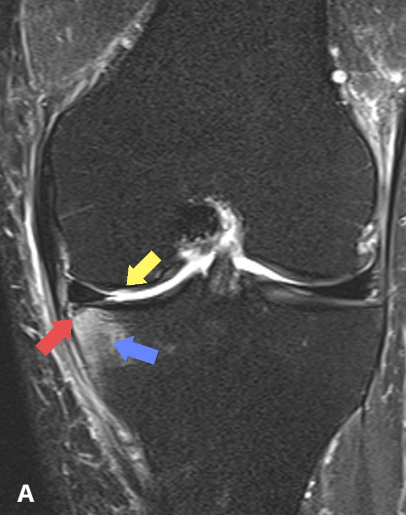
An MRI was performed demonstrating a medial meniscal tear at the posterior tibial root attachment, with a concurrent medial tibial plateau subchondral insufficiency fracture accompanied by moderate marrow oedema. There was no depression of the subchondral plate. Articular cartilage wear over the weightbearing medial femoral condyle of 50-75% depth.
However, on further questioning, the patient also symptoms further distally in the calf. This prompted further imaging down the distal lower leg.
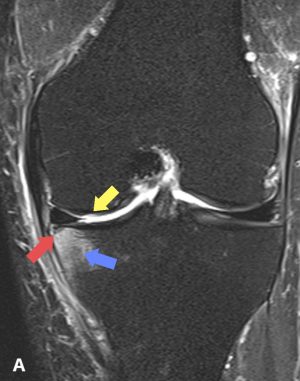
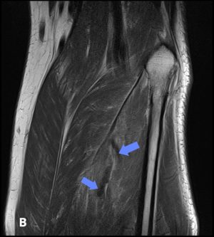
Figure A: Coronal T2 Fat saturated image of the knee demonstrating marrow oedema (blue arrow) and a subtle linear fracture (red arrow). Cartilage wear in the medial femoral condyle is demonstrated as well (yellow arrow)
Figure B: Coronal PD in the lower leg further distal demonstrating an unusual linear band like low signal in the calf muscles indicating a DVT (blue arrows).
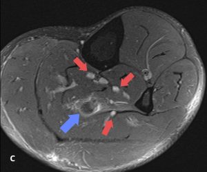
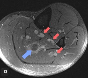
Figure C,D: Axial T2 fat saturated images showing a swollen and hypointense/dark appearance to one of the soleal veins in the calf with surrounding oedema (blue arrows) Note the presence of other deep veins appearing hyperintense/bright (red arrows)
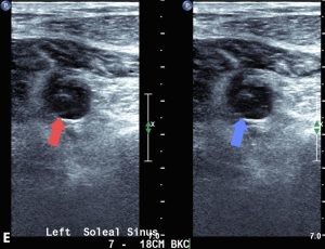
Figure E: Ultrasound confirming presence of DVT in soleal vein. This is an axial image with the image on the left without pressure on the calf (red arrow), and the image on the right during compression of the calf showing a filling defect in the vein, and non-compressibility (blue arrow)
Pathology
In this location, there is hypointense enlargement of the soleal deep veins with surrounding soft tissue oedema, in comparison to the surrounding deep veins in the calf which demonstrate T2 hyperintensity. This was suspicious for a deep venous thrombosis (DVT) and was confirmed on duplex ultrasound.
Comments
This case illustrates the importance of a thorough clinical examination and history in close correlation with imaging to detect a common pathology which is rarely diagnosed on MRI, namely deep venous thrombosis.
Although MRI features are suspicious for DVT in this case, the gold standard for detection of deep venous thrombosis is ultrasound, and this was required to be confident with the diagnosis.